Live Cell Imaging Services

Next Generation Cellular and Organotypic Assays
Senior Principal Scientist
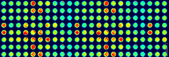
Live cell imaging methods use calcium dye to lead the cells and to be able to image the calcium inside the cells. In our laboratories, we use two different types of equipment:
Each of them offers certain benefits:
- a high spacial resolution (Zeiss),
- confocal (Zeiss),
- low cell numbers (Hamamatsu),
- higher throughput (Hamamatsu; 96/384 well),
- recording at physiological temperature 37°C,
- high-speed parallelized recording (Hamamatsu; up to 120 Hz).
Applications
- Safety profiling (seizure effects on neurons, arrhythmia on cardiac cells),
- compound MoA,
- intracellular Ca2+ handling,
- subcellular Ca2+ handling.
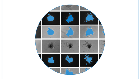
We use different models for studies that require live cell imaging approaches. Rat/mouse cortex, chicken cardiomyocytes, hiPSC-derived neurons with or without astrocytes, hiPSC-derived cardiomyocytes are a few we have to mention. In case such studies might help you with your scientific project, let us know.
Publications
-
Shen N, Knopf A, Westendorf C, Kraushaar U, Riedl J, Bauer H, Pöschel S, Layland S.L, Holeiter M, Knolle S, Brauchle E, Nsair A, Hinderer S, Schenke-Layland K. Steps toward Maturation of Embryonic Stem Cell-Derived Cardiomyocytes by Defined Physical Signals. Stem Cell Reports 2017 | Contact an author.
- Viero C, Kraushaar U, Ruppenthal S, Kaestner L, Lipp P. A Primary Culture System for Ssustained Expression of a Calcium Sensor in Preserved Adult Rat Ventricular Myocytes. Cell Calcium 2008 | Contact an author.
More Services
NMI Technologie Transfer GmbH
Markwiesenstraße 55, 72770 Reutlingen
Tel.: +49 7121 51530-0
Mail:
info@nmi-tt.de
NMI on Social Media
Follow us now


