Zeiss Cell Explorer Services

Next Generation Cellular and Organotypic Assays
Senior Principal Scientist
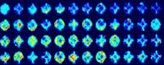
With attached Yokogawa spinning disk for rapid confocal imaging offers subcellular resolution with sampling frequencies up to 80 Hz.
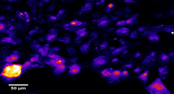
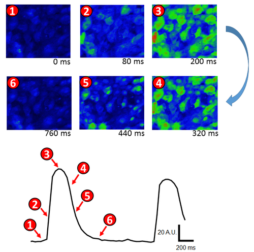
Confocal Ca2+ imaging of cultivated, spontaneously beating cardiomyocytes using the Zeiss Cell Observer system. The top part of the picture below: Time series of false-color coded Ca2+ dependent fluorescence of Cal520-AM loaded cardiomyocytes. Excitation leads to a transient intracellular Ca2+ increase, visualized by a color shift from blue via green to red. Bottom: Fluorescence intensity-over time trace of above-shown recording. Numbers indicate the time points of the images.
NMI Technologie Transfer GmbH
Markwiesenstraße 55, 72770 Reutlingen
Tel.: +49 7121 51530-0
Mail:
info@nmi-tt.de
NMI on Social Media
Follow us now


